.jpg)
The "visual window" of a blood bag, namely the transparent area on the bag, is primarily designed to allow medical staff to directly observe the state of the blood without opening the bag, ensuring blood quality and safety, and thus guaranteeing patient transfusion safety.
I. Key Points for Observing Blood State Through the Transparent Area
1. Observing Blood Color
Normal Condition: The color of normal blood varies depending on its oxygen content. Arterial blood has a high oxygen content and is bright red; venous blood has a low oxygen content and is dark red. In a blood bag, whole blood that has undergone anticoagulation treatment usually appears uniformly dark red. This is because red blood cells make up a large proportion of the blood and are in a relatively quiescent state, resulting in a lower oxygen content.
Abnormal Conditions and Analysis
Too Light in Color: If the blood color is significantly lighter than normal, it may indicate a decrease in the number of red blood cells, such as in cases of hemolysis. During hemolysis, red blood cells rupture, releasing hemoglobin into the plasma, causing the blood to lighten in color. The plasma may also appear pale red or pink. This situation may be caused by mechanical damage, excessively high or low temperatures, or bacterial contamination during blood collection, storage, or transportation. 1. **Excessively Dark or Black Blood Color:** Excessively dark or black blood may indicate the presence of methemoglobin or other abnormal substances. In methemoglobinemia, the increased methemoglobin content in the blood darkens its color. Additionally, prolonged storage or exposure to certain chemicals can also cause abnormal blood color.
2. **Observing Blood Layering:**
**Normal Condition:** After a certain period of settling in a blood bag, blood will separate into layers. The upper layer is pale yellow plasma, mainly containing water, protein, electrolytes, and nutrients; the lower layer is dark red red blood cells, possibly with a thin layer of white blood cells and platelets in between, called the white blood cell and platelet buffer layer. This layering is a normal physical characteristic of blood, indicating relatively stable blood composition.
**Abnormal Conditions and Analysis:**
**Indistinct or Absent Layering:** If blood layering is indistinct or absent, it may be due to violent shaking during collection or storage, causing red blood cells to break down and the plasma to mix with the red blood cells. This situation can affect blood quality and transfusion efficacy because broken red blood cells release intracellular substances, potentially causing transfusion reactions.
Abnormal stratification, such as the presence of flocculent material or sediment: The appearance of flocculent material or sediment may be due to fibrinogen precipitation, bacterial contamination, or immune complex formation in the blood. Fibrinogen plays an important role in blood clotting; when blood is affected by certain factors, fibrinogen may precipitate and form flocculent material. Bacterial contamination can lead to blood deterioration, producing sediment and an off-odor. Immune complex formation may be related to transfusion-related immune responses.
3. Observe for blood clots
Normal condition: In a qualified anticoagulant blood bag, blood should remain in a homogeneous liquid state without clot formation. This is because the anticoagulant prevents blood clotting and maintains blood fluidity.
Abnormal condition and analysis: If clots are found in the blood, it indicates that the anticoagulant may be ineffective, or there may have been improper operation during blood collection, such as an incorrect anticoagulant-to-blood ratio or excessively rapid collection leading to localized blood clotting. Transfusing blood containing clots can cause blood vessel blockage, leading to severe transfusion reactions and even endangering the patient's life.
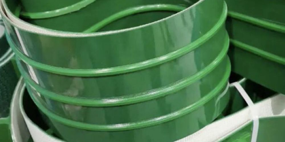
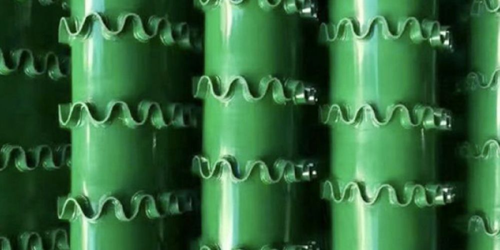
I. Different types of cleats and sidewall designs play unique roles in conveyor belt systems 1…
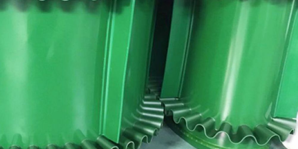
The sidewall design of sidewall-cleated conveyor belts enhances conveying efficiency in several ways: 1. **Increased Material…
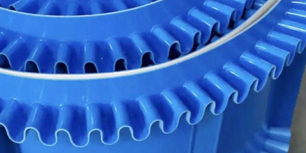
Skirted baffle conveyor belts are a special type of conveyor belt that combines the functions of…
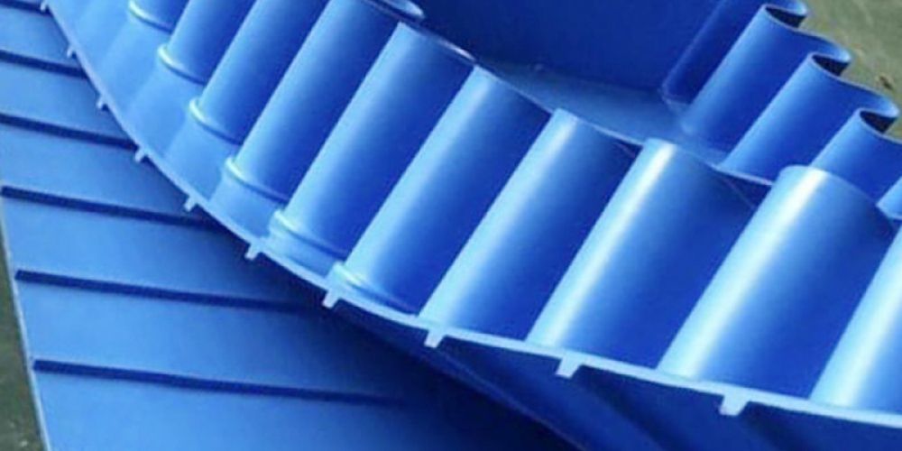
Polyurethane (PU) conveyor belts possess numerous advantages, including excellent wear resistance, oil resistance, corrosion resistance, non-toxicity…
The safety of conveyor belt materials in the food packaging industry can be ensured through several…
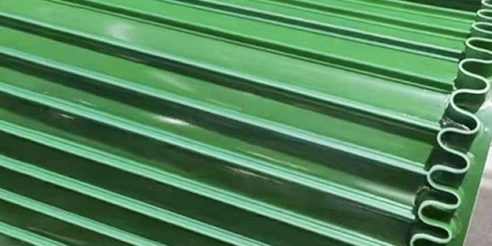
Lightweight conveyor belts are a type of conveyor belt made of relatively light materials. They come…